middle ear anatomy ct
Circle of Willis Anatomy. 9 Pictures about Circle of Willis Anatomy : The Middle Ear and Mastoid | Radiology Key, “If a Tree Falls – The Ear” and also Magnetic Resonance Imaging Of Inner Ear.
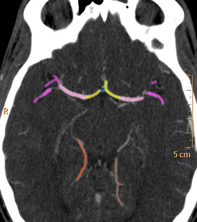
Circle Of Willis Anatomy
 www.radioanatomie.com
willis m2 cerebral segment circle m1 artery anatomie polygone anatomy middle anterior couleur a1 chargement des
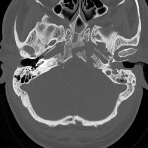
Skull Base Osteomyelitis | Image | Radiopaedia.org
 radiopaedia.org
osteomyelitis radiopaedia
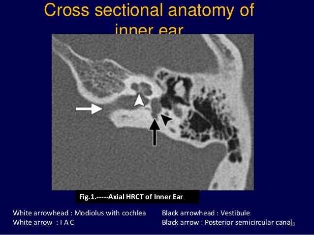
Temporal Bone (Middle Ear, Cochlea, Vestibular System) | Radiology Key
 radiologykey.com
cochlea vestibular
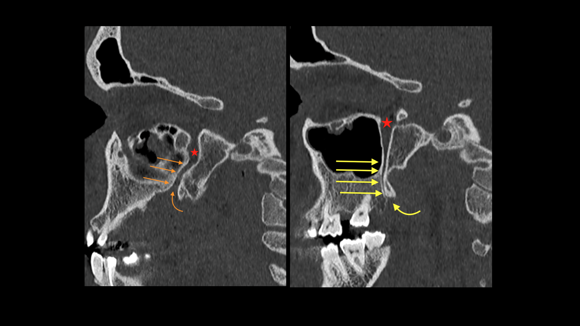
EPOS™ - C-2562
 epos.myesr.org
palatine canal lesser sagital foramen hrct ppf epos 2562 ecr2017 myesr esr
Facial Nerve Anatomy - Labeled CT | Image | Radiopaedia.org
 www.radiopaedia.org
nerve facial ct anatomy bone radiopaedia labeled axial window
The Middle Ear And Mastoid | Radiology Key
 radiologykey.com
mastoid ear middle
Magnetic Resonance Imaging Of Inner Ear
 www.slideshare.net
resonance hrct vestibular aqueduct transverse
“If A Tree Falls – The Ear”
 outlanderanatomy.com
ear bone temporal mastoid cells air middle anatomy section parts coronal showing bones falls tree fig muscles
Anatomia Rocca TC - CT Temporal Bone Anatomy
 www.slideshare.net
ct anatomy temporal bone tc anatomia ear rocca head bones 3d google inner mastoide facial radiology fossa del slideshare imaging
Temporal bone (middle ear, cochlea, vestibular system). Skull base osteomyelitis. Ear bone temporal mastoid cells air middle anatomy section parts coronal showing bones falls tree fig muscles